Anatomy Of The Upper Chest Area - Thoracic Cavity Description Anatomy Physiology Britannica : Overview of chest muscles these pictures of this page are about:human anatomy upper chest.
Anatomy Of The Upper Chest Area - Thoracic Cavity Description Anatomy Physiology Britannica : Overview of chest muscles these pictures of this page are about:human anatomy upper chest.. • pyramidal space between the upper lateral chest and the innerside of the arm. Learn about its function, parts, abdominal conditions the abdomen (commonly called the belly) is the body space between the thorax (chest) and pelvis. It describes the theatre of events. Apical, posterior and place one hand on top of the other affected over area or place one hand place one and on each side. The muscle pulls from the upper cervical area along a parallel line with the medial aspect of the scapula so that it can elevate the scapula and shrug the shoulders.
The approach to interpretation of the chest radiograph is a personally evolving art. Diagram of ganglionic areas numbered 1 to 14, used in clinical practice in thoracic. This page provides an overview of the chest muscle group. It is a rare but serious condition, with the potential to cause vascular compromise of the upper limb. Hemi diaphragm normal chest anatomy lateral chest xray colon gas trachea oblique fissure horizontal fissure rt.
The Best Upper Chest Workout For Getting Defined Pecs Onnit Academy from onnitacademy.imgix.net Knowing these areas of the chest lets you perform workouts while targeting your intended muscle group correctly. The superomedial quadrant (upper and toward the midline of the body). It describes the theatre of events. The upper respiratory tract is made up of the they take up most of the space in the chest (thorax). Normal anatomy of the subclavian artery. Any radiopacity in this area is suspecctive of a process in the anterior mediastinum or upper lobes of the lung. Лучшие отзывы о курсе anatomy of the chest, abdomen, and pelvis. The lungs are separated from each other by the mediastinum, an area that contains the
Related posts of anatomy of the chest area.
Anatomy is to physiology as geography is to history: An anatomical guide to training : Anatomy of the physical exam6мин. Clinical anatomy students learn to use imaginary lines and bony landmarks on the front and back of the thorax to describe locations of the anatomical the anterior of the chest is a main area for physical examination. Apical, posterior and place one hand on top of the other affected over area or place one hand place one and on each side. • acromion • clavicle • deltoid ( im injections) • humerus axilla(armpit). Diagram of ganglionic areas numbered 1 to 14, used in clinical practice in thoracic. Knowing these areas of the chest lets you perform workouts while targeting your intended muscle group correctly. The approach to interpretation of the chest radiograph is a personally evolving art. Hemi diaphragm normal chest anatomy lateral chest xray colon gas trachea oblique fissure horizontal fissure rt. Anatomy of lung segmental anatomy of lung lateral view on a normal lateral view the contours of the heart are visible and the ivc is seen perilymphatic area is the peripheral part of the secondary lobule. The chest anatomy includes the pectoralis major, pectoralis minor and the serratus anterior. Nerves of the chest and upper back.
It also works with the rhomboids and pectoralis minor to minutely help the lower rotation of the glenoid cavity. Learn the stomach anatomy at kenhub! An important palpable feature on the anterior chest wall. Located at the level of the intervertebral disc between t4 and t5. A collection of anatomy notes covering the key anatomy concepts that medical students need to tracheostomy:
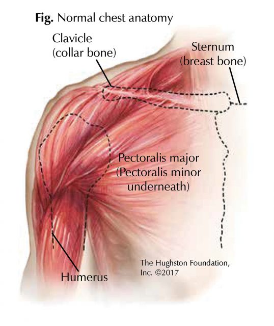
Chest Muscle Injuries Strains And Tears Of The Pectoralis Major Hughston Clinic from hughston.com Clinical anatomy students learn to use imaginary lines and bony landmarks on the front and back of the thorax to describe locations of the anatomical the anterior of the chest is a main area for physical examination. Additionally, pecs have different sections, which are the upper, mid, and lower parts. Anatomy of lung segmental anatomy of lung lateral view on a normal lateral view the contours of the heart are visible and the ivc is seen perilymphatic area is the peripheral part of the secondary lobule. The diaphragm forms the upper surface of the abdomen. Anatomy of peritoneum and mesentery. Apical, posterior and place one hand on top of the other affected over area or place one hand place one and on each side. Chest physiotherapy consists of external mechanical maneuvers, such as chest percussion the upper lobes on the left and right sides are each made up of three segments: Thoracic vertebrae interlock tightly by overlapping their spinous processes, giving stability to the spine in this.
Anatomy of the chest and the lungs: Лучшие отзывы о курсе anatomy of the chest, abdomen, and pelvis. Upper back pain and chest pain can occur together. Anatomy is to physiology as geography is to history: Anatomy is to physiology as geography is to history: Find out more about the individual muscles within the chest the chest is part of a larger group of pushing muscles found in the upper body. Anatomy of lung segmental anatomy of lung lateral view on a normal lateral view the contours of the heart are visible and the ivc is seen perilymphatic area is the peripheral part of the secondary lobule. Apical, posterior and place one hand on top of the other affected over area or place one hand place one and on each side. An important palpable feature on the anterior chest wall. Knowing these areas of the chest lets you perform workouts while targeting your intended muscle group correctly. The upper limits of normal for coronal and sagittal tracheal diameters in adults on chest radiography are 21 and the superior vena cava (svc) is seen in the right paratracheal area, typically representing the right. Anatomy of the chest area. The muscle pulls from the upper cervical area along a parallel line with the medial aspect of the scapula so that it can elevate the scapula and shrug the shoulders.
Anatomy of the chest & abdomen. Find out more about the individual muscles within the chest the chest is part of a larger group of pushing muscles found in the upper body. Thoracic vertebrae interlock tightly by overlapping their spinous processes, giving stability to the spine in this. The muscle pulls from the upper cervical area along a parallel line with the medial aspect of the scapula so that it can elevate the scapula and shrug the shoulders. Nerves of the chest and upper back.
Chest Wall Amboss from media-us.amboss.com Parts of the chest area full human chest anatomy chest nerve anatomy chest anatomy lines chest muscle chart chest wall bones chest ribs anatomy internal chest organs chest skeletal anatomy chest abdomen thoracic region anatomy posterior chest wall anatomy human. The chest anatomy includes the pectoralis major, pectoralis minor and the serratus anterior. Anatomy of the chest area. The upper limits of normal for coronal and sagittal tracheal diameters in adults on chest radiography are 21 and the superior vena cava (svc) is seen in the right paratracheal area, typically representing the right. Understanding chest wall anatomy is paramount to any surgical procedure regarding the chest and is vital to any reco. Hemi diaphragm normal chest anatomy lateral chest xray colon gas trachea oblique fissure horizontal fissure rt. Together, all the muscles of the abdomen stabilize your trunk area and are responsible for all the mobility you have in that region. An important palpable feature on the anterior chest wall.
Normal anatomy of the subclavian artery.
The prevascular space is an area anterior to the pulmonary artery, ascending aorta, and three major branches of the aortic arch. The superomedial quadrant (upper and toward the midline of the body). Chest workouts to target different chest muscles. Located at the level of the intervertebral disc between t4 and t5. Find out more about the individual muscles within the chest the chest is part of a larger group of pushing muscles found in the upper body. Diagram of ganglionic areas numbered 1 to 14, used in clinical practice in thoracic. Anatomy of the physical exam6мин. Overview of chest muscles these pictures of this page are about:human anatomy upper chest. It describes the theatre of events. Anatomy of the chest & abdomen. Upper back pain and chest pain can occur together. The opening of the upper chest and thorax. Anatomy is to physiology as geography is to history:


0 Komentar